📞Customer Service: +86 13248368268 📧servicecenter@suzhoufrank.com one year replacement and warranty!

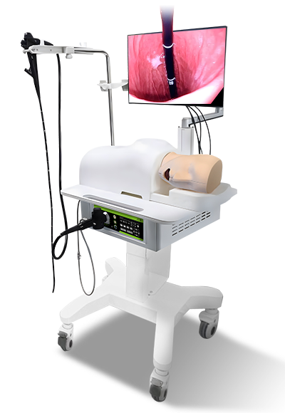
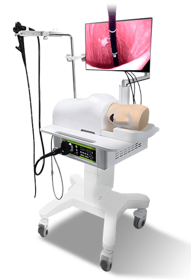
Endoscopy Simulator
ERCP Surgery Training Model
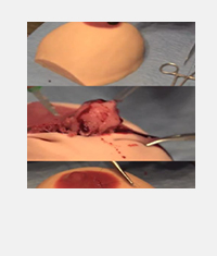
The Endoscopic Submucosal Dissection (ESD) Training Model is a medical simulation tool featuring 1:1 human upper gastrointestinal anatomical structure via 3D printing, using modified ex vivo porcine stomach/esophagus as training materials. It supports high-difficulty procedures like ESD, STER, and POEM, with replaceable components for realistic and repeated skill practice.
Brand: FESTAK
Product Type: Endoscopic Intervention Training Model
Warranty: 12 Month
whatsapp & Wechat: +86 15900838626
Anatomical Accuracy & Core Design
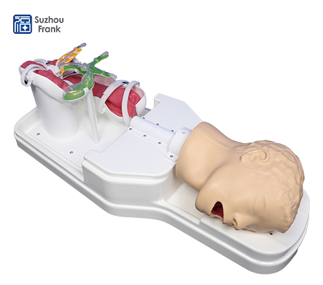
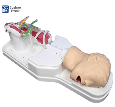
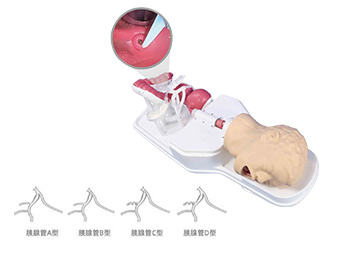
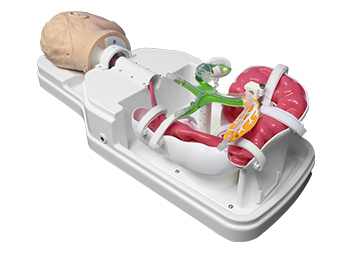
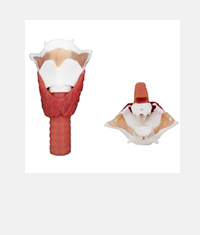
Built with 3D printing technology, the model replicates a 1:1 human upper gastrointestinal cavity (oral cavity, pharynx, esophagus, stomach). The esophagus-stomach transition uses soft material to enhance endoscope operability, ensuring alignment with real clinical anatomy.Components & Replaceable Parts
Key components include oral cavity, pharynx, esophagus, stomach body, base, and electrode connecting wires. Replaceable parts cover silicone esophagus, modified ex vivo porcine esophagus/stomach pieces, enabling sustained training after wear.Supported Procedures & Training Value
It facilitates training for high-difficulty endoscopic procedures: Endoscopic Submucosal Dissection (ESD), Submucosal Tunneling Endoscopic Resection (STER), and Peroral Endoscopic Myotomy (POEM). It also allows safe practice of perforation scenarios to understand treatment risks.
Advantage
Reusable, reducing costs
1:1 Reproduction of Human Body Data
Excellent Layering
Easy to operate
Easy to carry
Different modules for choice

Gallery


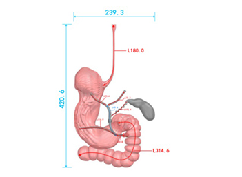
Parameter
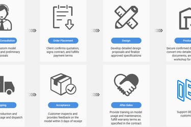
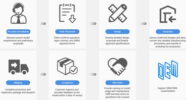
Model Customization Process


Client confirms quotation, signs contract, and fulfills payment terms


Develop detailed design proposals and finalize approved specifications
Secure confirmed designs and data, convert into detailed manufacturing documents, and transfer to workshop for production

Complete production and inspection, package and dispatch


Customer inspects and provides feedback on the model within 3 days of receipt


Provide training on model usage and maintenance, fulfill warranty terms as specified in the contract


Support OEM/ODM customization
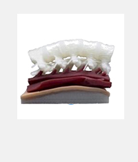
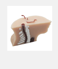
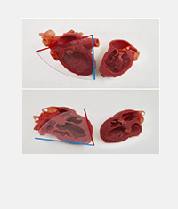
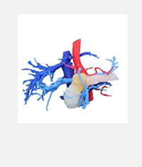
Client Cases

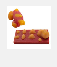
Breast tumour excision training model
Partial nephrectomy
Cystoscopy Training Model
ERCP Training Model
Lobectomy Training Model
Endoscopy Training Torso Model
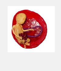
Obstetrics Model 1
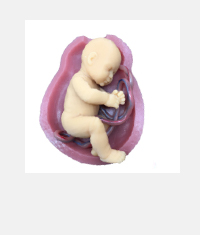
Obstetrics Model 2
Radiofrequency Ablation Model


Gastroesophageal Reflux Model
Tracheal Intubation Training Model
Model of pyeloplasty

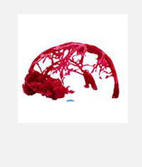

Cerebrovascular Tumour Model
Lateral Cerebral Puncture Drainage Model

Prostate Implant Training Model

Spinal anaesthesia puncture model

Neck anaesthesia Puncture model

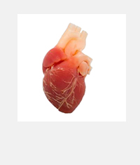
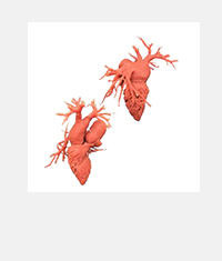
Cardiovascular Model

Tracheal Intubation Training Model

Mammary lesion excision

Varicose Vein Closure Training Model

Congenital Heart Disease Imaging Model


Pancreatic tumour model
TEE Cardiac ultrasound
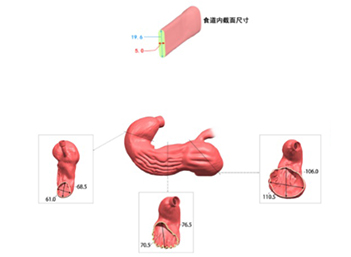
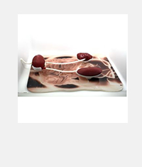
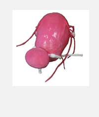
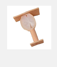
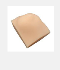
This 3D-printed ESD model uses modified ex vivo porcine stomach pieces, offering a realistic feel of human stomach walls for endoscopic submucosal dissection training.
3D Printed ESD Training Model with Ex Vivo Porcine Stomach
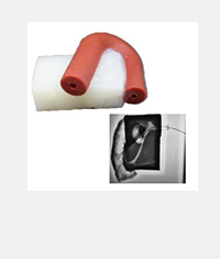
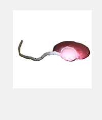
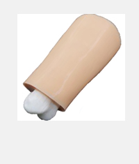
Featuring replaceable silicone esophagus and porcine tissue parts, this model supports repeated practice of ESD, STER, and POEM procedures for digestive endoscopy skills.
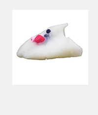
With a 1:1 anatomical upper gastrointestinal structure, this model is tailored for training high-difficulty endoscopic procedures like ESD and POEM, enhancing clinical operability.
Replaceable Component ESD/STER Endoscopic Training Model
1:1 Upper Gastrointestinal ESD Training Model for High-Difficulty Procedures
Frequently Asked Questions about ERCP Surgery Training Model
What’s the Upper Digestive Tract Endoscopy Training Model composed of?
Training high-difficulty endoscopic procedures like ESD, STER, and POEM.
What materials are used for training?
Modified ex vivo porcine stomach and esophagus pieces.
Does it have replaceable parts?
Yes, including silicone esophagus and porcine tissue components.
What anatomical structure does it replicate?
1:1 human upper gastrointestinal cavity (oral cavity, pharynx, esophagus, stomach).
Can it simulate perforation scenarios?
Yes, it allows safe practice of perforation to recognize treatment risks.
Is the esophagus-stomach transition easy for endoscopy?
Yes, the soft material at the transition enhances endoscope operability.
© 2025. All rights reserved.
About Us
Introduction
Development
Cooperation
Service
Main Products
Medical Grade Monitor
No 15, Jinyang road KunshanSuzhou, Jiangsu, China